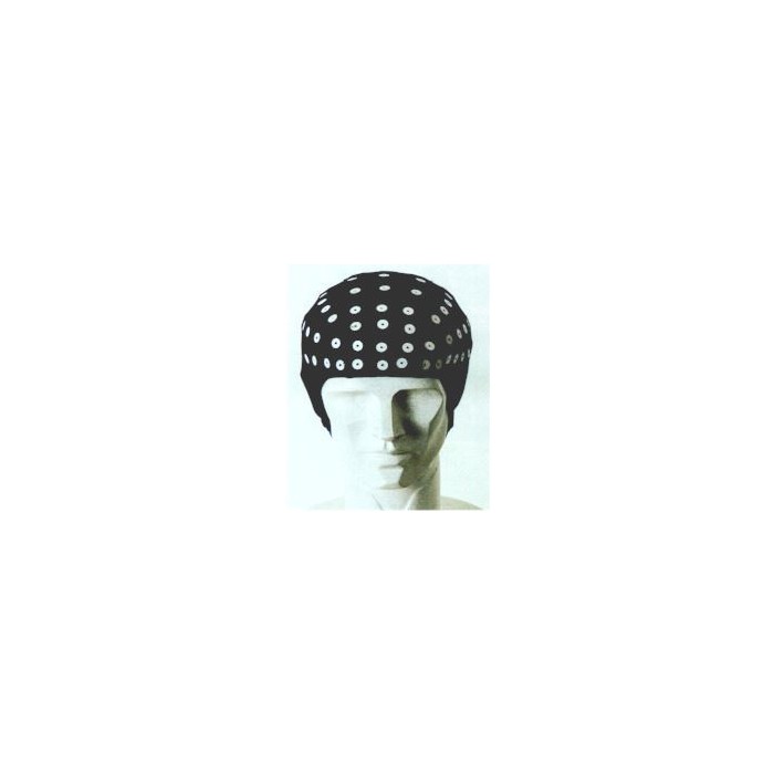
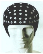
Special 128-channel EEG Cap from Electro-Cap (ECI). Caps come pre-assembled with 128 EEG electrodes positioned to the international standard 10-10 system. Different Electro-Cap sizes available. One 128-channel Electro-Cap plus one Electrode Board Adaptor (EBA) per pack.
This special 128-channel EEG cap is manufactured by ECI (Electro-Cap International). The fabric is an elastic spandex-type fabric. These EEG caps come pre-assembled with recessed EEG electrodes attached directly to the cap's fabric. The position of these electrodes is listed below.
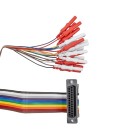
These special 128-channel Electro-Caps were originally specifically designed to work with EEG systems (amplifiers) from Neuroscan (E1-NSC128). This 128-channel Electro-Cap terminates in four 37-pin D-Sub connector. Four Electrode Board Adaptors (EBA) are also supplied which convert the 37-pin D-Sub connectors to DIN 1.5mm touch-proof (TP) sockets (female connectors). This allows this 128-channel Electro-Cap to be connected to any EEG system with touch-proof connectors.
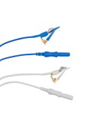
This 128-channel Electro-Cap is also special in that it includes additional electrodes for linked-ears (LE) and EOG channels (vertical and horizontal). These are accessible through so-called drops (Samtec sockets).
Electro-Caps are available in different sizes to fit adults, adolescents and children. The size medium (M) fits 65% of adolescents and adults. Smaller cap sizes will fit primarily children. Extra small caps fit most children from 9 months to 2 years of age. Sizes in age are approximate; measure the subject head with an ECI measuring tape to determine the suitable cap size.
Electro-Caps are used in various applications, including EEG, EP, LTM (Long-Term Monitoring), PSG (Polysomnography), Ambulatory, Telemetry, and even animal research.
The tips of the embedded electrodes can be either tin or Ag/AgCl. Check the specifications of your amplifier before selecting the appropriate electrode tip material. If you're not sure, the standard electrodes provided are tin electrodes.
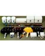
One special 128-channel Electro-Cap (ECI) EEG Cap per pack. Included with this 128-channel Electro-Cap are also two Electrode Board Adaptors (EBA) terminating all EEG channels in DIN 1.5mm touch-proof (TP) female connectors.
Electrode Position Layout
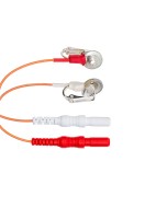
The 128 electrodes of this 128-channel Electro-Cap are positioned according to the international standard 10-10 system. Ground (GND) is located between FPz and Fz. This cap also features six drops for connecting electrodes to measure horizontal EOG (HEOG) and vertical EOG (VEOG). It is also equipped with ear-clip electrodes for using A1 and A2 as linked-ear (LE) reference.
Technical Specifications
- Cap Material: Elastic spandex-type fabric
- Size: 34cm - 62cm head circumference
- Number of Channels: 128
- Electrodes: Recessed EEG electrodes (pre-assembled)
- Applications: for EEG, EP, EEG for LTM, PSG and other EEG applications
- Packaging: 1 x 64-channel Electro-Cap (ECI) EEG Cap + 4 x EBAs per pack
Application note
- Connect the Electro-Cap to the electrode board adapter.
- Place the body harness around the chest and under the armpits of the subject.
- Fasten the velcro straps together making sure the straps are in front and centered on the chest.
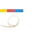
- Measured the circumference of the head with a special color code tap measure, one inch above the nasion and one inch above the inion. The color-coded tape will indicate the color of the proper cap size.
- Clip the earclip electrode to the subject’s earlobe.
- Using a syringe, inject a small amount of Electro-Gel into the disc cavity of the earclip electrodes. Rock the syringe back and forth using moderate pressure. This will abrade the skin and lower the impedance.
- To ensure proper placement of the electrodes, measure the distance of the nasion and inion to calculate the location of Fpz (10% of the distance between nasion and inion) and mark the location with a marking pen.
- Measure the circumference of the subject’s head to determine the location of the Fp1 and Fp2 (5% of the circumference left and right of Fpz and mark these locations with a marking pen.
- Place sponge disks over the Fp1 and Fp2 marks. These will absorb perspiration and reduce motion to the electrodes.
- Place the Electro-Cap on the subject’s head.
- Set the Fp1 and Fp2 into the sponge disks on the forehead.
- Continue to work the Electro-Cap on from front to back until it is tight on the head.
- Secure the cap straps to the body harness by crisscrossing straps and snapping them into place.
- Double-check that the straps have been pulled tight and secured. This will reduce movement artefacts during the EEG examination.
- Work each electrode onto the scalp to move excess hair from beneath the electrode mount. Make sure the Electro-Cap is centered on the head and fits properly.
- Connect all electrodes to your biosignal amplifier.
- Fill the syringe with 3-4cc of Electro-Gel.
- Place two fingers on the electrode holder and with the syringe, inject Electro-Gel while rocking the syringe back and forth using moderate pressure until a small amount of gel comes out of the hole. Wipe off the excess gel from the electrode.
- Check the impedances and ensure impedances are below 3 kOhms. If the impedance reads above 3 kOhms, the electrode must be refilled and the scalp re-abraded until the impedances improve. If the impedances stay above 3 kOhms, use a Quick Insert electrode which will bypass the electrode on the Electrode Board Adapter.
- Start the EEG recording.
-
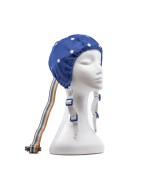 Electro-Cap (ECI) Standard 10-20 Single CapsAs low as $432.00
 -
 Infa-Cap (ECI) Infant Size Single Electro-CapsAs low as $391.20
 -
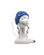 Electro-Cap (ECI) Lexicor with Ear DropsAs low as $501.60
 -
 Electro-Cap (ECI) Empty Cap (No Holders)As low as $264.00




 Australia & New Zealand
  Canada
  European Union (EU)
  France
  Germany
  Japan
Japan  Switzerland
  USA
USA  International